Take note of the incoming virtual meeting “SpectrIm – a tool for the combined analysis of MR Spectroscopy and Imaging” presented by Dr. Johannes Slotboom and Nuno Pedrosa de Barros, from the swiss Support Center for Advanced Neuroimaging (SCAN, Inselspital, University of Bern). This meeting is part of a series of planned virtual meetings on the topics: preprocessing, quantification, and simulation software for MRS/MRSI.
SpectrIm 1.0 beta screentshot
This virtual meeting is organized by the ISMRM Study Group on MR Spectroscopy and it will be held on Thursday 23 February at 07:00 PST, 10:00 EST, 16:00 CET. The meeting will start with a short introduction by Dr. Slotboom followed by a software demonstration by Nuno Pedrosa de Barros.
Note (*): This activity was restricted to members of the ISMRM MR Spectroscopy Study Group and required prior registration. To register, attendees had to go to the meeting registration page at ISMRM website and the login information was sent to registered attendees on Wednesday, 22 February 2017.
The virtual meeting video and the PowerPoint presentation are available for download at the ISMRM virtual meetings archive. Beware that a regular member login is required to access these files.
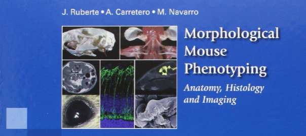
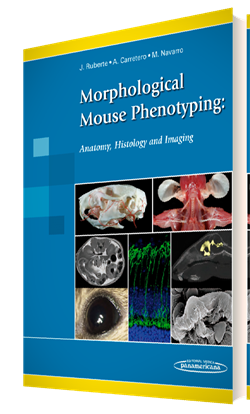
From the back cover of the book: Animal models of disease are fundamental in research to improve human health. The success of using genetically engineered mice to evaluate molecular disease hypotheses has encouraged the development of massive European and global projects making the mouse the most used animal model. Consequently, laboratory mouse populations are straining the housing capacity of pharmaceutical and biotechnology companies, as well as public research institutions. However, the scientific community often lacks sufficient expertise in morphological phenotyping to effectively characterize and validate these animal models.
Although the mouse displays fundamental morphological similarities to humans, a mouse is not a man. Here we present a complete and integrative description of normal mouse morphology. The main characteristics of this book are:
More than 2.200 original images have been specifically produced for this book in the Mouse Imaging Platform (Center for Animal Biotechnology and Gene Therapy, Universitat Autònoma de Barcelona).
These images show the anatomy, histology and cellular structure of mouse organs.
In addition, correlative X-ray, Computed Tomography, Magnetic Resonance (*) and Ultrasound images complete this integrative vision of the mouse morphology.
Classical anatomical techniques such as conventional dissection, skeletal preparations, vascular injections, as well as histological, immunohistochemical and electron microscopy techniques have been employed to characterize the mouse morphology.
This book, essentially an atlas, also contains explanatory diagrams and text that guides the reader through normal mouse anatomy, histology and imaging, and is aimed for mouse researchers as well as veterinarian and human pathologists.
(*) all Magnetic Resonance images in this book were acquired by Dr. Silvia Lope-Piedrafita at the at the NMR service (SeRMN) of Universitat Autònoma de Barcelona, in a 7 Tesla Bruker BioSpec 70/30USR spectrometer. SeRMN has been an active partner of the jMRUI community since many years.
jMRUI website uses cookies
Cookies are small text files held on your computer. We use them to improve your browsing experience of our site. By visiting our website you accept our use of cookies. For more information about cookies and how we use them, please visit our Our website uses cookies page.