Category Archives: General

morphological-mouse-phenotypig-featured-image

New atlas of mouse anatomy: from classical anatomical techniques to correlative X-ray, CT, MRI and Ultrasound images

If you use mice as animal model in research projects, make sure not to miss this new atlas of mouse anatomy:

Morphological Mouse Phenotyping: Anatomy, Histology and Imaging. 1st ed. Editorial Médica Panamericana S.A., 2016.

París, Jesús Ruberte, Romay Ana Carretero, and Beltrán Navarro. Morphological Mouse Phenotyping: Anatomy, Histology and Imaging. 1st ed. Editorial Médica Panamericana S.A., 2016.

From the back cover of the book: Animal models of disease are fundamental in research to improve human health. The success of using genetically engineered mice to evaluate molecular disease hypotheses has encouraged the development of massive European and global projects making the mouse the most used animal model. Consequently, laboratory mouse populations are straining the housing capacity of pharmaceutical and biotechnology companies, as well as public research institutions. However, the scientific community often lacks sufficient expertise in morphological phenotyping to effectively characterize and validate these animal models.

Although the mouse displays fundamental morphological similarities to humans, a mouse is not a man. Here we present a complete and integrative description of normal mouse morphology. The main characteristics of this book are:

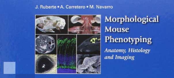
  • More than 2.200 original images have been specifically produced for this book in the Mouse Imaging Platform (Center for Animal Biotechnology and Gene Therapy, Universitat Autònoma de Barcelona).
  • These images show the anatomy, histology and cellular structure of mouse organs.
  • In addition, correlative X-ray, Computed Tomography, Magnetic Resonance (*) and Ultrasound images complete this integrative vision of the mouse morphology.
  • Classical anatomical techniques such as conventional dissection, skeletal preparations, vascular injections, as well as histological, immunohistochemical and electron microscopy techniques have been employed to characterize the mouse morphology.

This book, essentially an atlas, also contains explanatory diagrams and text that guides the reader through normal mouse anatomy, histology and imaging, and is aimed for mouse researchers as well as veterinarian and human pathologists.

The book is available from the publisher, from Amazon.com (check also your country website if available), and from other places.

(*) all Magnetic Resonance images in this book were acquired by Dr. Silvia Lope-Piedrafita at the  at the NMR service (SeRMN) of Universitat Autònoma de Barcelona, in a 7 Tesla Bruker BioSpec 70/30USR spectrometer. SeRMN has been an active partner of the jMRUI community since many years.

rssrss
FacebooktwitterlinkedinmailFacebooktwitterlinkedinmail
Teching at Paris (Wikipedia)

Non-commercial use of jMRUI in teaching-training activities

Few weeks ago we were contacted by Dr. Adam Liston, Course Director on the MSc in Advanced Neuroimaging at University College London, who wanted to know whether they could use the jMRUI software at the MSc in Advanced Neuroimaging for educational purposes. Although we knew from the very first moment that our answer would be “yes”, we decided to use his request to establish a set of general conditions the teaching and/or training activity would have to fulfil to be regarded as a non-commercial activity.

The use of the jMRUI software in teaching and/or training activities (e.g. postgraduate courses, workshops, etc.) does not constitute a commercial purpose as long as all these conditions are fulfilled:

  1. The jMRUI software is preferably installed on computers managed by the organising institution.
  2. If the jMRUI software is installed on student personal computers, students will be instructed that they must apply for a license if they want to keep the software after the course ends, and that otherwise they must delete it from their personal computers.
  3. The jMRUI software must be made freely available to the course attendees, and no fee can be charged to them for the distribution and/or the use of the software. Nevertheless, you can recoup the cost of the media: pendrive, cdrom, etc., used to distribute the software.
  4. The European Union research project currently funding the development of the jMRUI software must be acknowledge in the course brochure and/or website (if any) and in the teaching materials, at least in the part devoted to the jMRUI software. For that purpose you can use the text below or a similar sentence:

The current development of the jMRUI software is funded by TRANSACT – Transforming Magnetic Resonance Spectroscopy into a Clinical Tool (PITN-GA-2012-316679, http://www.transact-itn.eu), an EU-funded FP7-PEOPLE Marie Curie Initial Training Network running from 1st March 2013 till 28th February 2017. 

Last, although it is not a requirement, we would appreciate from you sending us a brief description of the course so that we can mention it on our TRANSACT project reports. Additionally, we may ask you for permission to publish a short post about the course on this jMRUI blog.

rssrss
FacebooktwitterlinkedinmailFacebooktwitterlinkedinmail