I am a senior NMR spectroscopist at Universitat Autonoma de Barcelona since 1994. That same year I learned about jMRUI and started collaborating with the project. Since 1997, I am responsible of the project web services and software distribution.
View all posts by Miquel →
We have two unfilled positions still available for Early Stage Researchers to work on the INSPiRE-MED European project.
We seek highly motivated and qualified individuals to fill the last two Early Stage Researcher three-years positions still available in the project. The successful candidates will contribute to the development of advanced biomedical research tools in the field of Magnetic Resonance Spectroscopy and Imaging, and its application to the clinical day-to-day practice.
The positions still available are:
ESR9: Glutamate neurotransmission in depression: combining receptor and metabolic information from simultaneous MR/PET. At The University of Manchester, United Kingdom. Job description – Apply
ESR12: A decision-support system based on MRSI data at 3T, for glioblastoma therapy response follow- up. At Consorcio Centro de Investigacion Biomédica en Red M.P. (CIBER), Barcelona, Spain. Job description – Apply
To apply to one or more of the available positions, follow the instructions at the bottom of their description document at the INSPiRE-MED website or click the “Where to apply” button in the description pages at the EURAXESS jobs portal.
Before applying, note that Early Stage Researchers are subject to the Eligibility and Mobility Rules:
Early-Stage Researchers shall, at the time of recruitment by the host organisation, be in the first four years (full-time equivalent research experience) of their research careers and have not been awarded a doctoral degree.
At the time of recruitment by the host organisation, researchers must not have resided or carried out their main activity (work, studies, etc.) in the country of their host organisation for more than 12 months in the 3 years immediately prior to the reference date.
We are recruiting 15 Early Stage Researchers to work on the INSPiRE-MED European project.
We seek highly motivated and qualified individuals to fill the 15 Early Stage Researcher three-years positions available in the project. The successful candidates will contribute to the development of advanced biomedical research tools in the field of Magnetic Resonance Spectroscopy and Imaging, and its application to the clinical day-to-day practice.
Before applying, note that Early Stage Researchers are subject to the Eligibility and Mobility Rules:
Early-Stage Researchers shall, at the time of recruitment by the host organisation, be in the first four years (full-time equivalent research experience) of their research careers and have not been awarded a doctoral degree.
At the time of recruitment by the host organisation, researchers must not have resided or carried out their main activity (work, studies, etc.) in the country of their host organisation for more than 12 months in the 3 years immediately prior to the reference date.
The available positions are:
ESR 1: Optimized sets of MRS scans for fingerprinting and dedicated parameter estimation by fitting or deep learning. At Universität Bern, Switzerland.
ESR 2: Maximize metabolic information in a multi-parametric study at ultra-high field, from pre-clinical to clinical validations. At École Polythechnique Fédérale de Lausanne, Switzerland.
ESR 3: Next generation of multinuclear MRS(I). At MR Code B.V., Utrecht, The Netherlands.
ESR 4: Implementation of high-resolution MRSI methods in a pre-clinical scanner, and optimization for brain longitudinal studies of therapy response in mice glioma models. At Universitat Autònoma de Barcelona, Spain.
ESR 5: Efficient quantum-mechanics based simulation of metabolite excitation for fingerprinting and iterative parameter estimation. At Ustav pristrojove techniky AVCR, v. v. i. (Institute of Scientific Instruments of the Czech Academy of Sciences), Brno, Czech Republic.
ESR 6: Quantification and cross-validation of metabolic biomarkers in preclinical disease models. At Katholieke Universiteit Leuven, Belgium.
ESR 7: Development of automatic pipelines for MRS(I)/PET data processing and machine learning methods for prognostic and classification in Multiple Sclerosis patients. At Université Claude Bernard-Lyon1, Lyon, France) –
ESR 8: Pathophysiology of Tourette syndrome: multimodal characterization of metabolic alterations. At Max-Planck-Gesellschaft Zur Forderung Der Wissenschaften Ev (Max Planck Institute for Human Cognitive and Brain Sciences), Leipzig, Germany.
ESR9: Glutamate neurotransmission in depression: combining receptor and metabolic information from simultaneous MR/PET. At The University of Manchester, United Kingdom.
ESR10: Integrated acquisition & post-processing of MR spectroscopic imaging (MRSI) data of the prostate. At Radboud University Medical Center, Nijmegen, The Netherlands.
ESR11: Automated nosologic imaging by fusing multiparametric MR information in longitudinal patient follow-up. At Katholieke Universiteit Leuven, Belgium.
ESR12: A decision-support system based on MRSI data at 3T, for glioblastoma therapy response follow- up. At Consorcio Centro de Investigacion Biomédica en Red M.P. (CIBER), Barcelona, Spain.
ESR13: Boosting the academic jMRUI with novel algorithms and data format standardization. At Ustav pristrojove techniky AVCR, v. v. i. (Institute of Scientific Instruments of the Czech Academy of Sciences), Brno, Czech Republic.
ESR14: Validation and CE documentation of a Magnetic Resonance Spectroscopy clinical pipeline as a medical device. At ICOMETRIX N.V., Leuven, Belgium.
ESR15: Development of robust, user-friendly analysis and reporting tools for automatic MRS(I) processing for use in a clinical setting. At Universitätsspital Bern, Switzerland.
To apply to one or more of the available positions, follow the instructions at the bottom of their description document at the INSPiRE-MED website or click the “Where to apply” button in the description pages at the EURAXESS jobs portal.
Integrating Magnetic Resonance Spectroscopy and Multimodal Imaging for Research and Education in MEDicine (INSPiRE-MED) is an European research project awarded in the call H2020-MSCA-ITN-2018, of the MSCA-ITN-ETN – European Training Networks, to a consortium of 12 academic partners and 9 industrial partners coordinated by Prof. Dominique Sappey-Marinier, of the Université Lyon-1 Claude-Bernard, Lyon, France.
Starting 1st of January 2019, the INSPIRE-MED Initial Training Network will investigate over the next four years the theoretical and practical aspects of in vivo Magnetic Resonance Spectroscopy (MRS) and Spectroscopic Imaging (MRSI) with applications in oncology and neurology.
MRS(I) is a unique non-invasive technique that provides insight on in-vivo metabolite content, an information that is highly relevant for the diagnosis and therapy follow-up in numerous disease models and patients. Despite such potential, clinical uptake of MRS(I) is lagging behind that of MRI and PET, mainly because of limited availability of efficient, robust and automatic software tools. Thus, INSPiRE-MED aims at establishing MRS(I) as an additional tool integrated into clinical routine imaging, but also extending its benefits with next-generation research methods. For this purpose, INSPiRE-MED will create novel MRS(I) methodology, integrate MRS(I) into multimodal MR/PET clinical metabolic imaging protocols, develop latest machine learning methods for data analysis and provide a clinical version of jMRUI, a worldwide unique data processing tool for MRS(I).
If you are a user of either the jMRUI software, the INTERPRET Decision Support System or the SpectraClassifier tool, we would like to know how you use these software tools and your opinion about them, so that we can keep improving our software to better serve your needs.
For that purpose, we ask you to participate in the TRANSACT-ITN final software survey by answering few questions about your training, your working environment, the data you process, and how you use and rate the software. Although most questions are mandatory, many are so trivial that you will answer them in a blink, and the full questionnaire should take you less than 15 minutes to complete, and less than 10 minutes if you only use the jMRUI software. The questionnaire has been tested with the most popular web browsers running on desktop computers and mobile phones and tablets, and you should be able to answer it from elsewhere.
The survey is brought to you by TRANSACT, the EU-funded FP7-PEOPLE Marie Curie Initial Training Network (ITN) project that has been fostering the development of the jMRUI software from March 1, 2013 to February 28, 2017. The aim of the project was to Transform Magnetic Resonance Spectroscopy into a Clinical Tool, and two of the main objectives of the project were:
to improve the usability of the jMRUI software in the clinics, and
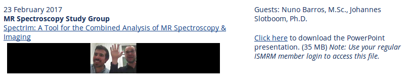
Take note of the incoming virtual meeting “SpectrIm – a tool for the combined analysis of MR Spectroscopy and Imaging” presented by Dr. Johannes Slotboom and Nuno Pedrosa de Barros, from the swiss Support Center for Advanced Neuroimaging (SCAN, Inselspital, University of Bern). This meeting is part of a series of planned virtual meetings on the topics: preprocessing, quantification, and simulation software for MRS/MRSI.
SpectrIm 1.0 beta screentshot
This virtual meeting is organized by the ISMRM Study Group on MR Spectroscopy and it will be held on Thursday 23 February at 07:00 PST, 10:00 EST, 16:00 CET. The meeting will start with a short introduction by Dr. Slotboom followed by a software demonstration by Nuno Pedrosa de Barros.
Note (*): This activity was restricted to members of the ISMRM MR Spectroscopy Study Group and required prior registration. To register, attendees had to go to the meeting registration page at ISMRM website and the login information was sent to registered attendees on Wednesday, 22 February 2017.
The virtual meeting video and the PowerPoint presentation are available for download at the ISMRM virtual meetings archive. Beware that a regular member login is required to access these files.
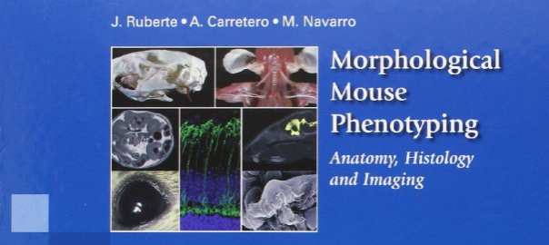
From the back cover of the book: Animal models of disease are fundamental in research to improve human health. The success of using genetically engineered mice to evaluate molecular disease hypotheses has encouraged the development of massive European and global projects making the mouse the most used animal model. Consequently, laboratory mouse populations are straining the housing capacity of pharmaceutical and biotechnology companies, as well as public research institutions. However, the scientific community often lacks sufficient expertise in morphological phenotyping to effectively characterize and validate these animal models.
Although the mouse displays fundamental morphological similarities to humans, a mouse is not a man. Here we present a complete and integrative description of normal mouse morphology. The main characteristics of this book are:
More than 2.200 original images have been specifically produced for this book in the Mouse Imaging Platform (Center for Animal Biotechnology and Gene Therapy, Universitat Autònoma de Barcelona).
These images show the anatomy, histology and cellular structure of mouse organs.
In addition, correlative X-ray, Computed Tomography, Magnetic Resonance (*) and Ultrasound images complete this integrative vision of the mouse morphology.
Classical anatomical techniques such as conventional dissection, skeletal preparations, vascular injections, as well as histological, immunohistochemical and electron microscopy techniques have been employed to characterize the mouse morphology.
This book, essentially an atlas, also contains explanatory diagrams and text that guides the reader through normal mouse anatomy, histology and imaging, and is aimed for mouse researchers as well as veterinarian and human pathologists.
(*) all Magnetic Resonance images in this book were acquired by Dr. Silvia Lope-Piedrafita at the at the NMR service (SeRMN) of Universitat Autònoma de Barcelona, in a 7 Tesla Bruker BioSpec 70/30USR spectrometer. SeRMN has been an active partner of the jMRUI community since many years.
Few weeks ago we were contacted by Dr. Adam Liston, Course Director on the MSc in Advanced Neuroimaging at University College London, who wanted to know whether they could use the jMRUI software at the MSc in Advanced Neuroimaging for educational purposes. Although we knew from the very first moment that our answer would be “yes”, we decided to use his request to establish a set of general conditions the teaching and/or training activity would have to fulfil to be regarded as a non-commercial activity.
The use of the jMRUI software in teaching and/or training activities (e.g. postgraduate courses, workshops, etc.) does not constitute a commercial purpose as long as all these conditions are fulfilled:
The jMRUI software is preferably installed on computers managed by the organising institution.
If the jMRUI software is installed on student personal computers, students will be instructed that they must apply for a license if they want to keep the software after the course ends, and that otherwise they must delete it from their personal computers.
The jMRUI software must be made freely available to the course attendees, and no fee can be charged to them for the distribution and/or the use of the software. Nevertheless, you can recoup the cost of the media: pendrive, cdrom, etc., used to distribute the software.
The European Union research project currently funding the development of the jMRUI software must be acknowledge in the course brochure and/or website (if any) and in the teaching materials, at least in the part devoted to the jMRUI software. For that purpose you can use the text below or a similar sentence:
The current development of the jMRUI software is funded by TRANSACT – Transforming Magnetic Resonance Spectroscopy into a Clinical Tool (PITN-GA-2012-316679, http://www.transact-itn.eu), an EU-funded FP7-PEOPLE Marie Curie Initial Training Network running from 1st March 2013 till 28th February 2017.
Last, although it is not a requirement, we would appreciate from you sending us a brief description of the course so that we can mention it on our TRANSACT project reports. Additionally, we may ask you for permission to publish a short post about the course on this jMRUI blog.
UCL Institute of Neurology (left) and the National Hospital for Neurology and Neurosurgery (right) in Queen Square, London (Source: Wikipedia).
The MSc in Advanced Neuroimaging is a multidisciplinary programme which aims to give students a strong working knowledge of neuroanatomy and an in-depth understanding of standard and advanced neuroimaging techniques for image acquisition, processing and analysis in the diagnosis, treatment and study of a full range of neurological diseases. During their time at Queen Square, students will have the opportunity to contribute to world-leading research and have access to cutting edge neuroimaging facilities.
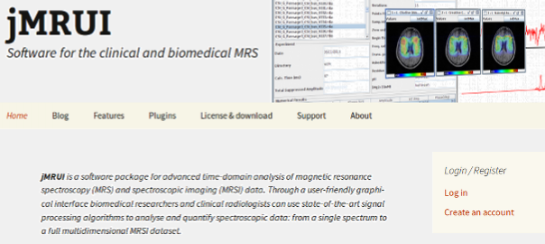
It has been a long while since we deployed the jMRUI website and after years of ageing it was intensely pleading for a thorough update. The work began under the FAST European Project, but it has finally come to light with the current TRANSACT European Project.
The new website runs on WordPress, a free and open source blogging and content-management software running on PHP and MySQL. According to Wikipedia, WordPress is used by more than 23.3% of the top 10 million websites as of January 2015, and it is the most popular blogging system in use on the Web at more than 60 million websites.
It is our hope that this new website will foster the development of a virtual community that will work towards spreading the use of MR spectroscopy in the pre-clinical and clinical worlds.
jMRUI website uses cookies
Cookies are small text files held on your computer. We use them to improve your browsing experience of our site. By visiting our website you accept our use of cookies. For more information about cookies and how we use them, please visit our Our website uses cookies page.